CT Reconstruction
Fibrocystic Breast Disease
I use the INVESALIUS program to convert CT scan data into obj. file.
Because I want to focus on the thoracic area, I only export the upper part of the body into ZBRUSH.

The original file contains many debris and holes that need to be fixed.

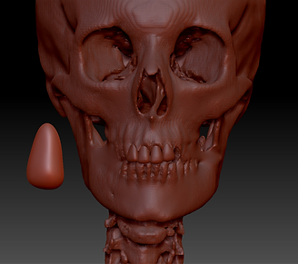
I started by filling the holes in the bone. It was challenging at the beginning, because it was my first time trying ZBRUSH. I spend few days to be familiar with brushes and tools.


After that, I sculped the teeth. I tried to check the anatomical accuracy of teeth with many references. I had planned to focus on the thorax, but I thought having half a transparent layer of the facial bone will make the final outcome look more realistic.

Then, I smooth the bones and skin of the model. I filled all the holes in skin and emphasised breast, as I might illustrate breast cancer or other disease which symptoms appear on the breast later.


To a point where all the necessary structures are fixed and repaired, I added the colour to bones and skin.


Then, I rendered the file in Keyshot. It was also my first attempt at Keyshot. I adjusted the lighting and added more texture to the model, and rendered it as 2D images in different positions.


I received comments from the critique section to draw more structure at the lower part of the breast. I also thought it looked better after I draw that part. The image looked more fulfilled and completed. I checked the anatomical accuracy and lighting with life drawing section in My Dundee.
I plan to illustrate fibrocystic breast disease, so I studied the structure around the breast. Because the model body shape is thick, I interpreted that she may have a high fat content in her body, so I draw a larger fat structure than the reference from the Netter Clinical Anatomy textbook (NETTER, F. H., HANSEN, J. T., & LAMBERT, D. R. (2005). Netter's clinical anatomy. Carlstadt, N.J., Icon Learning Systems.). I draw on other sides of the breast in the same model to show significant benign diseases. I tried to make the structure as clear as possible, and easy to understand for medical students. These process were done with photoshop.


Final outcome
I would like to focus on the clinical cases around the breast, so I have created an illustration of fibrocystic breast disease in Adobe illustrator program. The level of detail is based on Netter's clinical anatomy textbook. This content will be useful for medical students and those in related fields who study anatomy.
